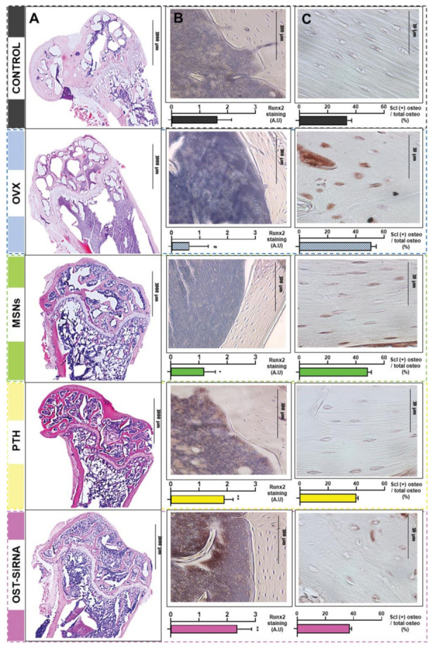
Figure 6.

Histological analysis and immunostaining in the femur of osteoporotic mice at 3 weeks post-treatment with mesoporous silica nanoparticles (MSNs) grafted with alendronate-modified poly(ethylene glycol) and poly(ethylene imine) (MSNs-PA@PEI), parathyroid hormone (PTH), and MSNs-PA@PEI loaded with osteostatin and sclerostin-encoding plasmid (OST-SiRNA), evidencing: representative micrographs of different femur histological sections after hematoxylin/eosin and Masson–Goldner trichrome staining (A); representative Runx2 immunostaining in mice femurs, revealed by the abundant positivity (brown stains) for the transcription factor in cells after PTH or OST-siRNA treatments (B); total and sclerostin-positive osteocytes in the cortical femur (C). Data are represented as mean ± standard error of mean of five independent mice (n = 5), and the statistical significance is indicated as # p < 0.001 vs. control, * p < 0.05 vs. ovariectomized mice (OVX), and ** p < 0.001 vs. OVX. See Ref. [302]. Reprinted from an open access source.
