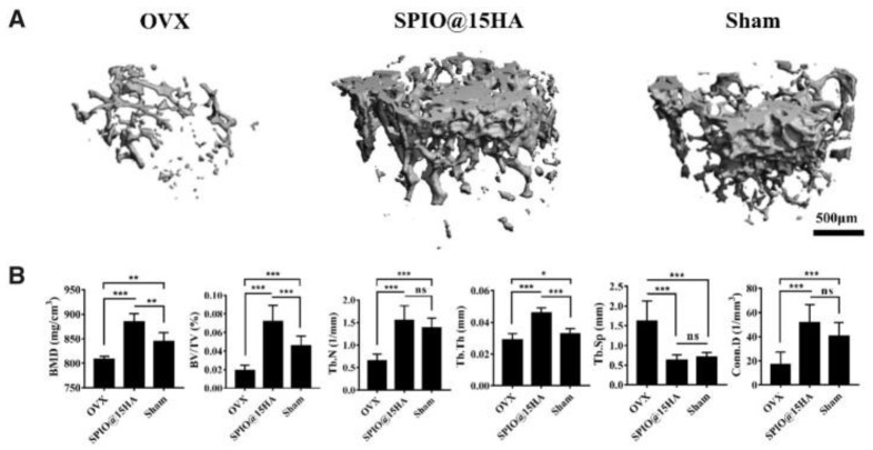
Figure 8.
Three-dimensional μ-CT reconstruction images of trabecular bone in ovariectomized mice (OVX), OVX treated with hydroxyapatite-coated superparamagnetic iron oxide nanocomposites (SPIO@15HA) and sham group (A), and trabecular bone mass parameters (B), evidenced after 3 months post-injection. BMD—bone mineral density, BV/TV—bone volume fractions, Tb.N—trabecular number, Tb.Th—trabecular thickness, Tb.Sp—trabecular spacing, Conn.D—connectivity density. Data are expressed as mean ± standard deviation of seven independent mice (n = 7), ns means no significance, and the statistical significance is indicated as * p < 0.05, ** p < 0.01, and *** p < 0.001. See Ref. [370]. Reprinted from an open access source.

