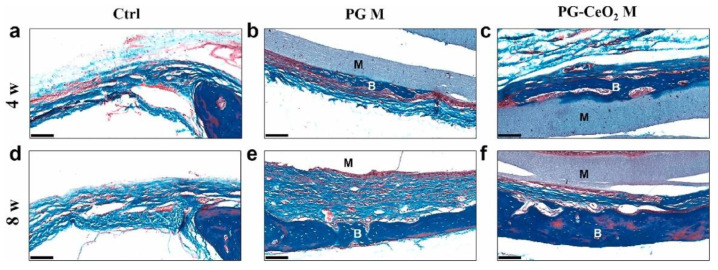
Figure 9.
Histological analysis of rat cranial defects treated with bare and nano-ceria-loaded polycaprolactone/gelatin membranes (PG M and PG-CeO2 M, respectively) for 4 and 8 weeks (w), evidenced by Masson’s trichrome staining. Control group (a,d), PG M group (b,e), and PG-CeO2 M group (c,f). M—membrane, B—bone, scale bar—100 μm. See Ref. [417]. Reprinted from an open access source.

