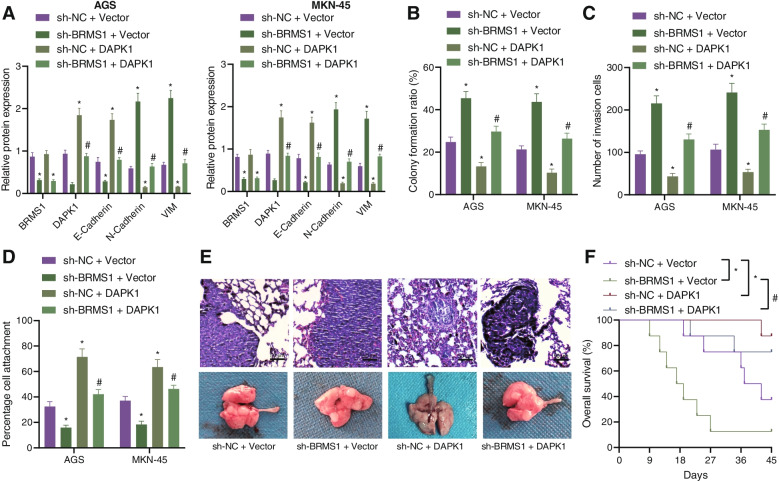
Fig. 6.
BRMS1 represses the proliferation and invasion of GC cells via DAPK1 enhancement. AGS and MKN-45 cells were treated with sh-BRMS1, DAPK1, or both. A Western blot analysis of BRMS1, DAPK1, E-cadherin, N-cadherin, and Vimentin proteins in cells. B Colony formation of AGS and MKN-45 cells. C Invasion of AGS and MKN-45 cells measured by Transwell assay. D Matrix adhesion ability of AGS and MKN-45 cells. E HE staining analysis of the lung tissues of mice treated with sh-BRMS1, DAPK1, or both (scale bar = 50 μm). F Survival curve of nude mice treated with sh-BRMS1, DAPK1, or both. Each experiment was conducted three times independently. n = 8 for mice following each treatment. * p < 0.05, compared with AGS and MKN-45 cells or mice treated with sh-NC + Vector. # p < 0.05, compared with AGS and MKN-45 cells or mice treated with sh-BRMS1 + Vector

