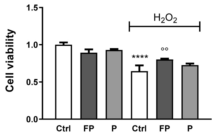
Figure 3.
HUVECs were plated in six-well plates (1.5 × 10⁵ cells/well) and treated with 10 μg/mL of FP and P for 24 h followed by treatment with H2O2 (300 µM) for 24 h. Cell viability was determined by Trypan Blue staining. Results are expressed as mean ± SD of at least three experiments. **** p < 0.0001 significantly different from the control (ctrl). °° p < 0.01, significantly different from H2O2-treated HUVECs. Ctrl (control, untreated cells); H2O2 (cells treated with hydrogen peroxide); FP (cells pretreated with fermented Pushgay); P (cells pretreated with Pushgay).

