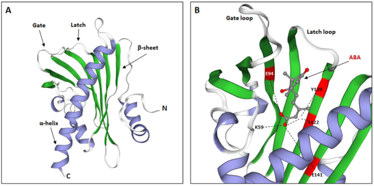
Figure 4.
ABA receptor structure. (A). Structure of the monomeric, apo-form of Festuca elata PYR1 (FePYR1), showing a secondary structure: α-helixes, β-sheets, and two main loops, involved in the ABA binding: latch and gate. (B). Structure of AtPYR1 binding pocket, highlighting main amino acids involved in the interaction with ABA residues: K59, E94, Y120, S122, and E141. (Modified from [71]).

