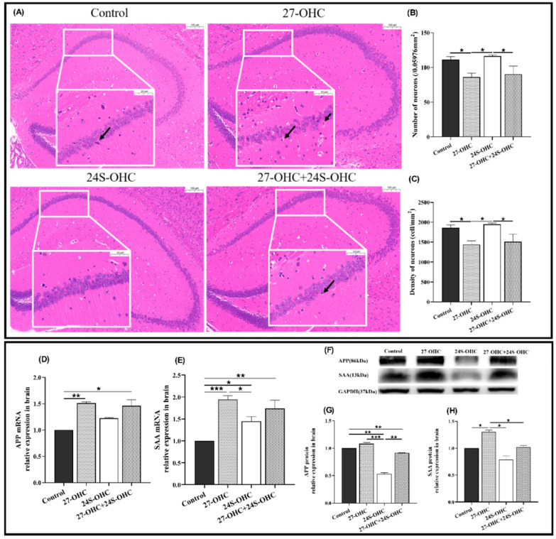
Figure 3.
Effects of oxysterols on brain pathology and the expression of amyloid precursor protein in the brain. (A) HE staining of the whole brain (n = 3, scale bar: 100 and 20 μm), black arrows: Nuclei pyknosis; (B) number of neurons (/0.05976 mm2) (n = 3, mean ± SEM); (C) density of neurons (cell/mm2) (n = 3, mean ± SEM); (D) APP mRNA (n = 7, median with range); (E) SAA mRNA (n = 7, mean ± SEM); (F) Western blot results of APP and SAA; (G) APP protein (n = 7, mean ± SEM); (H) SAA protein (n = 7, mean ± SEM). *: p < 0.05, **: p < 0.01, ***: p < 0.001.

