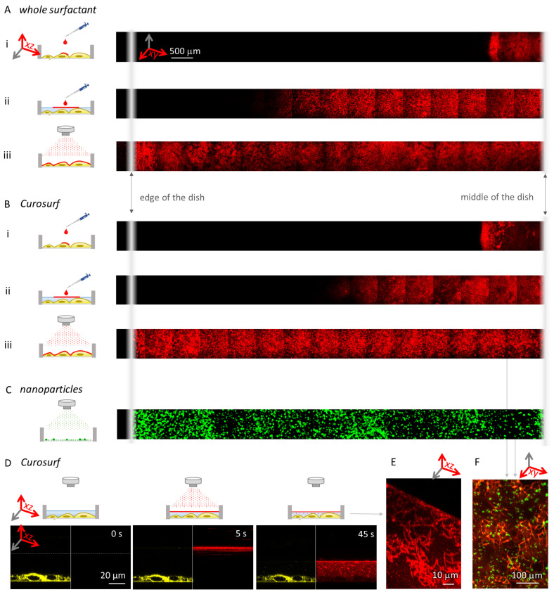
Figure 3.
The deposition of nebulized and pipetted surfactant. A total of 10 monolayers of fluorescently labelled (A) whole surfactant and (B) Curosurf were administered onto cells using different methods. As shown in the top-view panoramas, in both cases (i) surfactant, pipetted onto cells with removed medium (simulating ALI conditions), spreads only over a fraction of the sample, (ii) surfactant pipetted onto cells slightly covered with cell medium spreads further, but still does not cover the entire sample, and (iii) nebulized surfactant evenly covers the entire sample (fluorescence intensities are reported in Figure S3). Note that the intensities are not comparable between measurements, and the dark vertical lines are the consequence of uneven illumination. (C) Nebulization of fluorescently labelled TiO nanotubes (green) evenly deposits the material over the sample—shown here in a top-view panorama for nebulization directly onto a glass surface. Note that, due to the low signal, the image was filtered using a Gauss filter, and the intensity was scaled from 0 to 3 counts. (D) A time series of nebulization of 10 monolayers of Curosurf (red, fluorescently labelled with STAR RED-DPPE) onto lung alveolar cells (green, labelled with CellMask Orange), (see Figure S2 for complete time series). (E) A side-view of the surfactant structure just below the surface after nebulizing 100 monolayers of surfactant (red) onto submerged cells. The surface of the medium is tilted due to capillary effects near the edge of the dish. (F) A fluorescence top-view micrograph of 10 monolayers of nebulized surfactant (red) and subsequently nebulized 1:1 surface dose of nanomaterial (green) onto lung alveolar cells.

