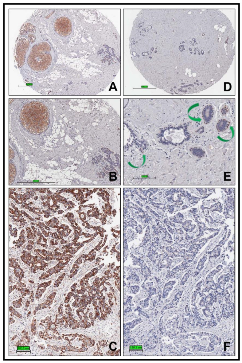
Figure 1.
Plg-RKT is highly expressed in human ductal carcinoma in situ and invasive ductal carcinoma. A core from a patient with ductal carcinoma in situ was stained with anti-Plg-RKT mAb using paraffin immunocytochemistry (A) and enlarged in (B). Cores from the same patient showing invasive ductal carcinoma (C) and from a 60-year-old healthy female control subject ((D) and enlarged in panel (E)) were stained in the same way. Green arrows in panel E indicate cells with macrophage morphology that also stain with anti-Plg-RKT mAb. A specificity control in which the core shown in Panel C was stained with anti-Plg-RKT mAb that had been absorbed with the Plg-RKT peptide used for immunization is shown in Panel F. Original magnifications, ×100 (A,D), ×200 (B) to ×400 (C,E,F).

