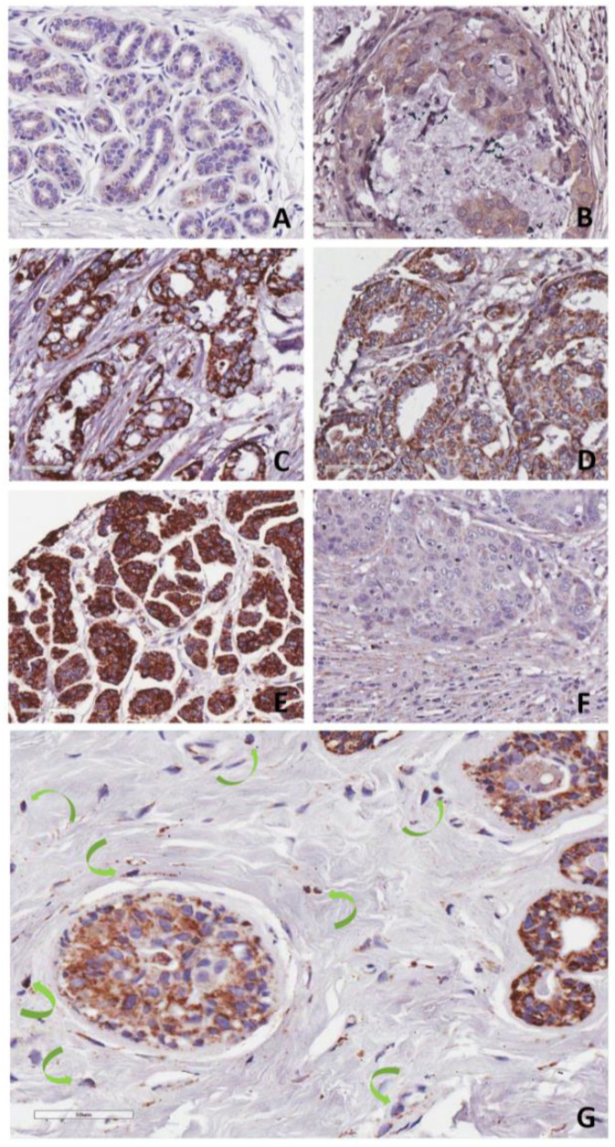
Figure 2.
Plg-RKT expression in human mammary tissues. (A) Ductal epithelial cells in normal breast with light granular Plg-RKT staining, (B) ductal carcinoma in situ with faint to moderate staining, (C) invasive, LN-positive HR-positive breast tumor, (D) invasive, HR-positive tumors with distant metastases show moderate and (E) strong granular Plg-RKT staining. (F) Invasive, LN-positive triple-negative breast tumor exhibits very fine granular Plg-RKT staining of tumor cells and faint to moderate staining of the reactive stroma. (G) Ductal carcinoma in situ showing staining of TAMs (identified by morphology and indicated with green arrows). Plg-RKT by IHC using anti-Plg-RKT monoclonal antibody 7H1. Shown are representative tissues from the CDP breast cancer progression TMA. Original magnifications, ×100 (A,E), ×200 (C,D,F) to ×400 (B,G).

