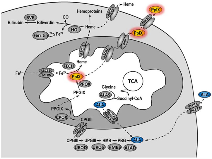
Figure 1.
Heme metabolism pathway. ALA is normally synthetized in the mitochondrial matrix by combining glycine with succinyl-CoA in a reaction catalyzed by ALA synthase (ALAS). After passing into the cytoplasm, ALA is converted into coproporphyrinogen III (CPGIII) as a result of numerous changes. Exogenously administered 5-ALA is delivered to the cytoplasm of cells by peptide transporter 1 and 2 (PEPT1 and PEPT2). CPGIII is transported to the intermembrane space of mitochondria where it is converted by coproporphyrinogen oxidase (CPOX) to protoporphyrinogen IX (PPGIX) and then in the mitochondrial matrix to PpIX by protoporphyrinogen oxidase (PPOX). Next, ferrochelatase (FECH) leads to the formation of heme by incorporating iron into PpIX. PpIX and heme can be released from the cell via the ABCG2 transporter. Heme in the cell is used to create hemoproteins or it is degraded by heme oxygenase (HO), leading to the release of biliverdin, CO, and Fe2+. Iron ions may be bound and stored by ferritin. Biliverdin is converted to bilirubin by biliverdin reductase (BVR). ABCB10—ABC subfamily B member 10; ALAD—5-aminolevulinic acid synthase dehydratase; HMB—hydroxymethylbilane; MFRN—mitoferrin; PBG—porphobilinogen; PBGD—porphobilinogen deaminase; PBR—peripheral-type benzodiazepine receptor; SLC25A38—solute carrier family member 38; TCA—tricarboxylic acid; UPGIII—uroporphyrinogen III; UROD—uroporphyrinogen decarboxylase; UROS—uroporphyrinogen III synthase.

