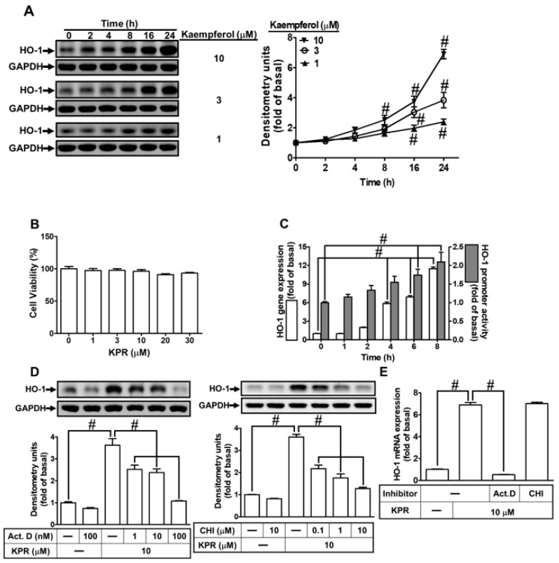
Figure 3.
KPR induces HO-1 expression in HPAEpiCs. (A) HPAEpiCs were treated with various concentrations of KPR for the indicated time intervals. The protein expression of HO-1 was determined by Western blot using GAPDH as a loading control. (B) Cells were treated with various concentrations of KPR for 24 h and the cell viability was examined by an XTT kit. (C) HPAEpiCs were treated with KPR (10 μM) for the indicated time intervals. The HO-1 mRNA expression and promoter activity were analyzed by real-time PCR and promoter activity assay kit, respectively. (D) HPAEpiCs were preincubated with various concentrations of either Act. D or CHI for 1 h and then incubated with vehicle or KPR (10 μM) for 16 h. The levels of HO-1 protein expression were determined by Western blot using GAPDH as a loading control. (E) Cells were pretreated with/without 100 nM Act. D or 10 μM CHI for 1 h, and then incubated with DMSO or KPR (10 μM) for 6 h. The level of HO-1 mRNA expression was analyzed by real-time PCR. Data are expressed as mean ± SEM (n = 5), analyzed with one way ANOVA and Dunnett’s post hoc test. # p < 0.01, as compared with the cells exposed to vehicle alone; or significantly different as indicated.

