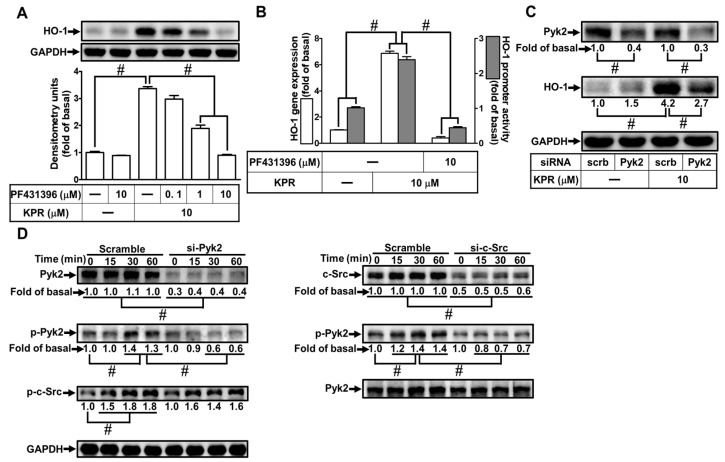
Figure 6.
Pyk2 involves in KPR-induced HO-1 expression. (A) HPAEpiCs were pretreated with various concentrations of PF431396 for 1 h and then incubated with DMSO or KPR (10 μM) for 16 h. The protein expression of HO-1 was determined by Western blot using GAPDH as a loading control. (B) Cells were pretreated with or without PF431396 (10 μM) for 1 h and then incubated with DMSO or KPR (10 μM) for the indicated time intervals. The HO-1 mRNA expression and promoter activity were analyzed by real-time PCR (6 h) and promoter activity assay (8 h), respectively. (C) Cells were transfected with scrambled (scrb) or Pyk2 siRNA, and then incubated with KPR (10 μM) for 16 h. The protein level of HO-1, Pyk2, and GAPDH was determined by Western blot. (D) Cells were transfected with c-Src or Pyk2 siRNA, and then incubated with KPR (10 μM) for the indicated time intervals. The levels of phospho-Pyk2, total c-Src, and total Pyk2 were determined by Western blot. Data are expressed as mean ± SEM (n = 5), analyzed with one way ANOVA and Dunnett’s post hoc test. # p < 0.01, as compared with the cells exposed to vehicle alone; or significantly different as indicated.

