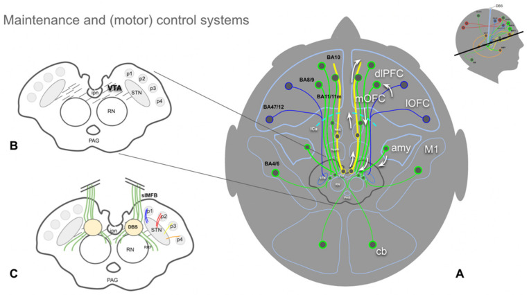
Figure 4.
Overview of the MFB (MAINTENANCE) and part of the (motor) control system. (A), yellow fibers represent the imMFB and, therefore, the principal DA projection to cortical and subcortical structures. Green fibers represent cortical and cerebellar feedback projections to the VTA (Glu). Blue fibers visualize (part of) the motor control system, with projections from the lOFC and dlPFC to the STN (Glu, hyperdirect pathway). (B), midbrain topographic overview. The hatched region (VTA) is the principal origin of the DA fibers (A10 and medial A9 cell groups). (C), same as (B) but with slMFB fibers (green) included. Descending fibers diverge in front of the red nucleus (RN) to reach the VTA and raphe as well as the lateral tegmentum (RRF, retro-rubral field, DA group A8). (Legend: amy, amygdala; ipn, interpeduncular nucleus; DBS, deep brain stimulation electrode; STN, subthalamic nucleus; PAG, periaqueductal gray; p1, frontopontine tract; p2, corticobulbar tract; p3, corticospinal tract; p4; occipito-temporo-pontine tract; cb, cerebellum; OFC, orbitofrontal cortex; mOFC, medial and central OFC; lOFC, lateral OFC; dlPFC, dorsolateral prefrontal cortex; M1, primary motor cortex; BA, Brodmann’s area.)

