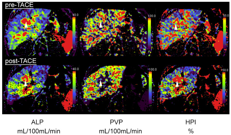
Figure 2.
A 64-year-old patient with central HCC and incomplete embolization after transarterial chemoembolization (TACE). Pre-TACE images (top row) show increased arterial-liver perfusion (ALP, left, white arrowhead), slightly reduced portal-venous perfusion (PVP, middle) and increased hepatic-perfusion index (HPI, right) compared to normal liver tissue. Post-TACE images (bottom row) reveal a slight decrease in ALP (left), indicative of residual-tumor tissue.

