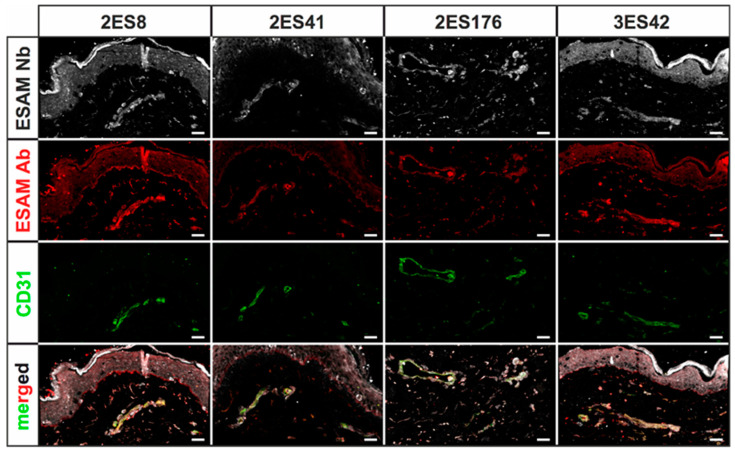
Figure 3.
Immunofluorescence stainings of human skin cryosections using hESAM-specific Nbs. Representative images of 2D immunofluorescence stainings of 5 µm cryosections of human skin specimens stained with the respective Nbs (white), a commercially available hESAM antibody (red), and CD31 (green), an antibody visualizing blood vessels. Stained antigens are indicated next to each panel, the Nb clone used is indicated above each panel. Scale bars = 50 µm.

