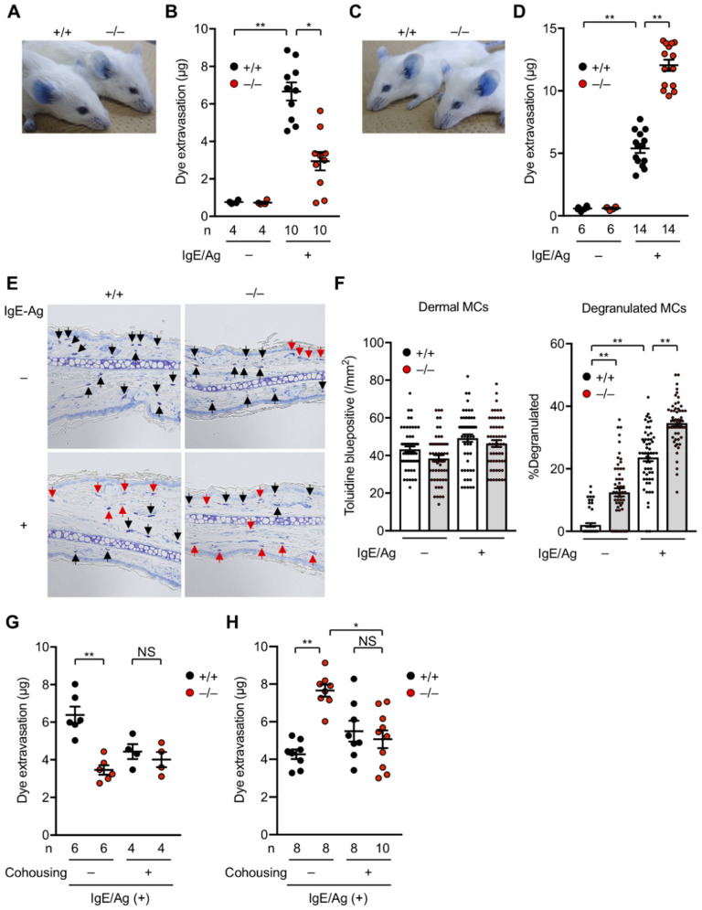
Figure 3.
Altered PCA responses in Pla2g2a−/− BALB/c mice under distinct housing conditions. Ears of Pla2g2a+/+ and Pla2g2a−/− mice (8–12 weeks of age, male) were sensitized by subcutaneous injection of anti-DNP IgE monoclonal antibody (30 ng) and then challenged by intravenous injection of a mixture of DNP-conjugated human serum albumin (60 µg) as an antigen (Ag) together with 1 mg Evans blue, as described previously [75]: (A–D) Representative photos of the ears (A,C) and quantification of dye extravasation (B,D) in IgE/Ag-treated (+) or -untreated (−) Pla2g2a+/+ and PLa2g2a−/− mice housed in the TMIMS (A,B) and UTokyo (C,D) facilities. (E,F) Histology of the skin (E) and quantification of total and degranulated mast cells (F) in IgE/Ag-treated or -untreated Pla2g2a+/+ and Pla2g2a−/− mice housed in the UTokyo facility. The ear pinnae were fixed with 10% (v/v) formalin, embedded in paraffin, sectioned (4-µm thickness), and stained with toluidine blue. A total of 55 views for each group (n = 5). Black and red arrows indicate non-degranulated and degranulated mast cells, respectively. Scale bar, 25 µm. (G,H) IgE/Ag-induced PCA reaction in Pla2g2a+/+ and Pla2g2a−/− mice with (+) or without (−) co-housing in the TMIMS (G) and UTokyo (H) facilities. Values are mean ± SEM. *, p < 0.05; **, p < 0.01; NS, not significant. Statistical analysis was performed using Graph Pad PRISM with Brown–Forsythe test and then Kruskal–Wallis and Dunn’s post hoc test (B,D,F,G,H). The numbers of mice used for the analysis are indicated in each panel.

