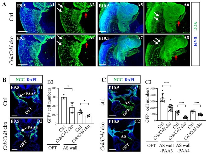
Figure 3.

Greatly reduced NCC localization in the aortic sac wall and OFT in Crk/Crkl cko embryos. (A) Sagittal view and the three-dimensional (3D) reconstruction of whole mount GFP (green) embryos. Slightly fewer migratory NCCs in streams in Crk/Crkl cko versus Wnt1-Cre/+; Crkf/+; Crklf/+ double heterozygous control embryos. White and red arrows in (A) indicate migrating NCC streams and cardiac OFT, respectively. Defective migrating NCC streams were shown in (A4, A8). N = 4 for each genotype. (B, C) Less NCCs in cardiac OFT and dorsal aortic sac wall regions in Crk/Crkl cko versus control embryos at E9.5 (B) and E10.5 (C) as shown by immunofluorescence (B1–2, C1–2). Quantification of NCC numbers in cardiac OFT and dorsal aortic sac wall regions in Crk/Crkl cko versus control embryos at E9.5 (B3) and E10.5 (C3). Each dot in graph represented one embryo (B3, C3). The graphs (B3, C3) were plotted with mean and standard deviation. Two-tailed Student’s t-test was used for the statistical analysis. *: 0.01 < P < 0.05. ****: P < 0.0001. The genotype of the control embryos in (A–C) is Wnt1-Cre/+; Crkf/+; Crklf/+. PAA3, third pharyngeal arch artery; AS, aortic sac. Scale bar in (A1–4, C1–2): 200 μm. Scale bar in (A5–8): 500 μm. Scale bar in (B1, B2): 100 μm.
