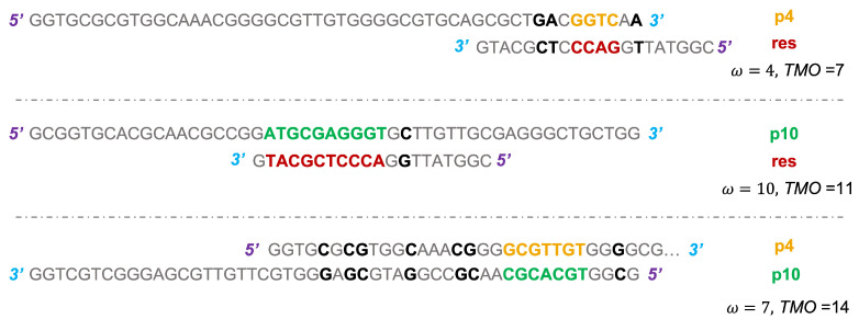
Figure 3.
Scheme of the binding between p4 and res (top), where the bases forming the MCO are in bold font and colored in gold (p4) and in firebrick (res). The other bases contributing to the TMO are in black color and bold font. The same applies for p10-res interaction (middle), where the bases of p10 involved in the formation of MCO are colored in green. The (bottom) panel shows the configuration for the formation of MCO between a p4 and a p10 strand, with the usual color scheme. The 5 and 3 ends are colored, respectively, in purple and cyan, highlighting the reciprocal orientation of strands.

