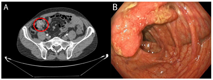
Figure 1.
(A) Concentric wall thickening of the ileocecal valve compatible with a neoplasia. Approximate measures: 2.8 × 3 × 2.8 cm (T × AP × L). No spiculations on the paracecal fat or regional adenopathy are observed. (B) Endoscopic view of tumour. Flat and over-raised lesion with central depression of at least 20 mm in the colonic wall, suggestive of adenocarcinoma.

