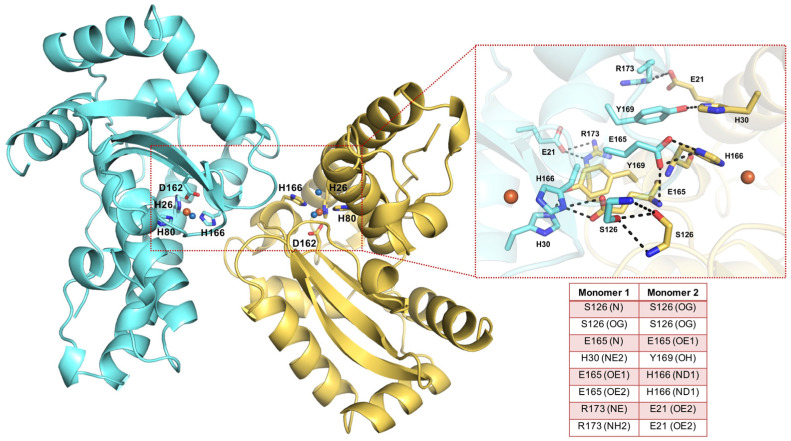
Figure 1.
Overview of SmSOD architecture and residues at the dimer interface. The X-ray structure of SmSOD (PDB 4YIP) is shown as a ribbon model, with the two monomers coloured in aquamarine and yellow. The active site residues (H26, H80, H166, D162) are shown as sticks; the metal ion and the coordinating water molecules are shown as orange and blue spheres, respectively. On the right side, a zoom-in of the residues composing the dimer interface in which H-bonds are depicted as black dashed lines and listed in the inserted table.

