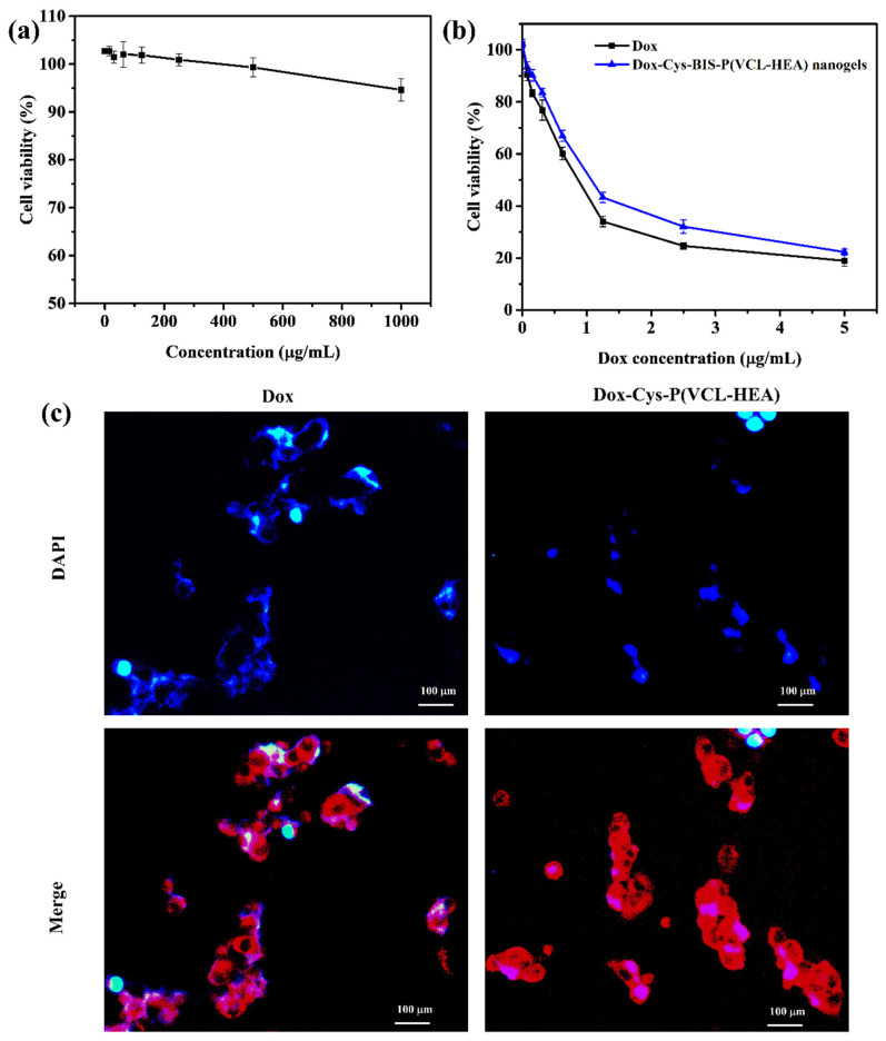
Figure 5.
(a) In vitro cytotoxicity of pure nanogels treated with CCDK-normal skin fibroblasts cells with different concentrations (0–1000 μg/mL) for 72 h incubation. (b) In vitro cytotoxicity of Dox-loaded Cys-BIS-P(VCL-HEA) nanogels treated with HepG2 cancer cells. (c) Fluorescence microscopy images of samples treated with HepG2 cells for 3 h incubation (DAPI (blue) staining for nucleus and red fluorescence represent the Dox or Dox-loaded nanogels).

