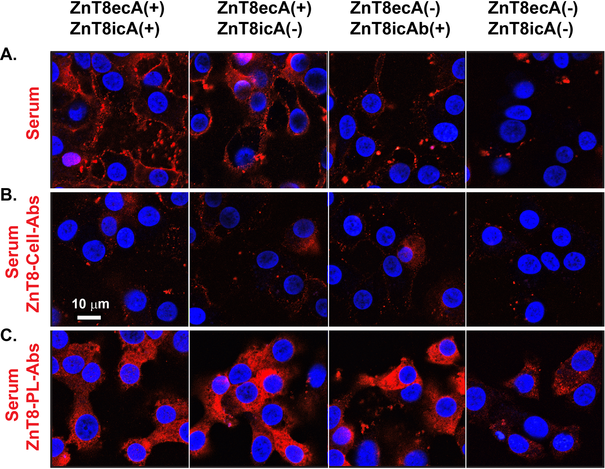
Fig. 3. Cell surface immunofluorescence staining of ZnT8ecA from sera of individual diabetic patients.

A. Human EndoC-βH1 cells were exposed to a human serum that was selected for ZnT8ecA and ZnT8icA positive or negative in various combinations as indicated. B-C. Identical sera were pre-absorbed by ZnT8-GFP on the surface of intact INS-1E cells (ZnT8-cell-Abs) or by purified human ZnT8 in proteoliposomes (ZnT8-PL-Abs) as indicated. Serum antibodies bound to the cell surface were visualized by confocal microscopy using a secondary antibody against human IgG (red) while cell nuclei were counterstained by DAPI (blue). Representative images are shown from immunofluorescence staining using two independent serum sets, each with 4 replicates.
