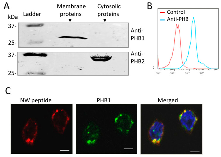
Figure 5.
Analysis of prohibitin cellular localization in monocytes. (A) Western blot analysis. Membrane and cytosolic proteins were prepared from monocytes and analyzed via Western blotting using monoclonal antibodies against PHB1 or PHB2. (B) A representative flow cytometry histogram showing the expression of PHB1 on the surface of monocytes. (C) PHB1 co-localizes with the NW peptide on the plasma membrane of monocytes. Monocytes were co-stained with the NW peptide and a mouse anti-prohibitin monoclonal antibody, followed by incubation with PE-streptavidin and anti-mouse IgG-FITC, and then stained with DAPI. Cells were mounted on slides, and confocal images were recorded. The data are representative of at least three independent experiments. Scale bar = 20 μm. PHB, Prohibitin; kDa, Kilodaltons.

