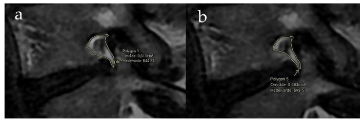
Figure 2.
MRI of a right L4–5 foramina without loading of the spine (a) and MRI of the same foramina acquired with axial loading (b). The measurement area is marked in both images and a reduction in the area was measured. The fat surrounding the nerve root is visually reduced in the MRI with loading.

