Figure 3.

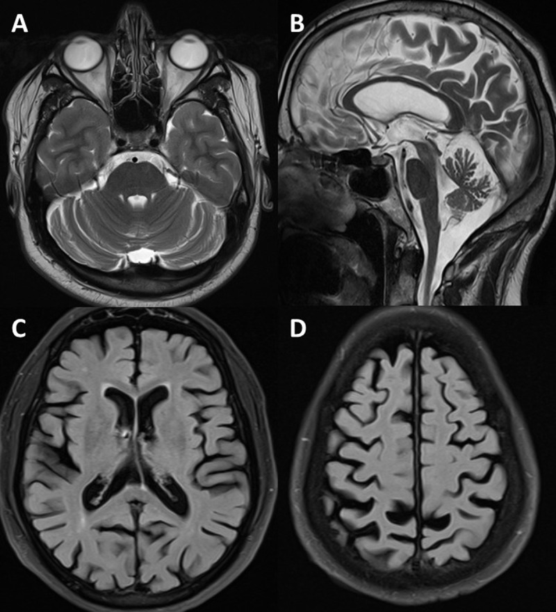
MRI brain showing cerebellar atrophy in (A) T2 axial and (B) mid-sagittal sections and cerebral atrophy in (C, D) FLAIR axial sections.

MRI brain showing cerebellar atrophy in (A) T2 axial and (B) mid-sagittal sections and cerebral atrophy in (C, D) FLAIR axial sections.