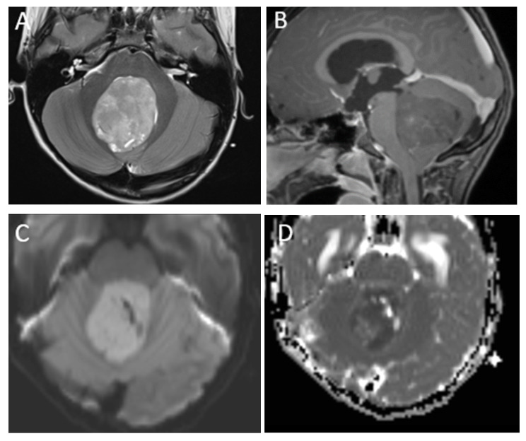
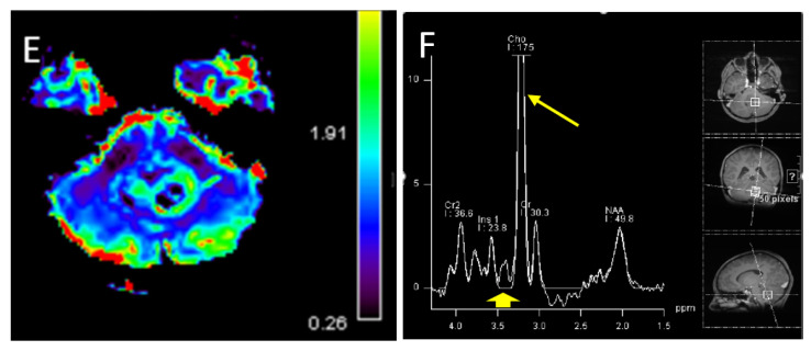
Figure 1.
A 7-year-old boy with medulloblastoma. Axial T2 (A), sagittal post-contrast T1 (B), images demonstrate a mildly heterogeneous midline posterior fossa mass. DWI (C) and ADC (D) imaging demonstrates diffusion restriction of the lesion relative to the adjacent cerebellar parenchyma, typical of these highly cellular neoplasms. Perfusion imaging (E) demonstrates increased relative cerebral blood volume within the lesion and MRS (F) demonstrates a high choline peak (long arrow) as well as a small taurine peak (short arrow).


