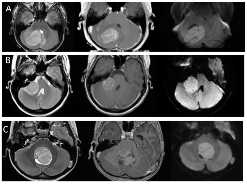
Figure 3.
Imaging appearances of molecular subtypes of medulloblastoma. Each row includes axial T2-weighted, axial post-contrast T1-weighted and axial diffusion-weighted images from left to right. (Row (A)): SHH. Imaging demonstrates an off-midline lesion with the classic cerebellar hemispheric location of these lesions. (Row (B)): WNT. Imaging demonstrates the classic CP angle location of this subtype, although many of these lesions may arise in the midline as well. (Row (C)): Group 3 and (Row (D)): Group 4. These tumors are classically located in the midline. Note the relative hypoenhancement of the group 4 tumor, which has been a described feature. All lesions demonstrate characteristic restricted diffusion.


