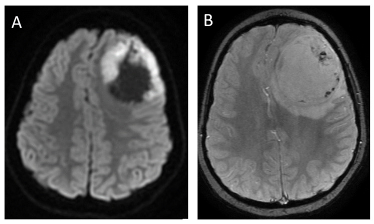
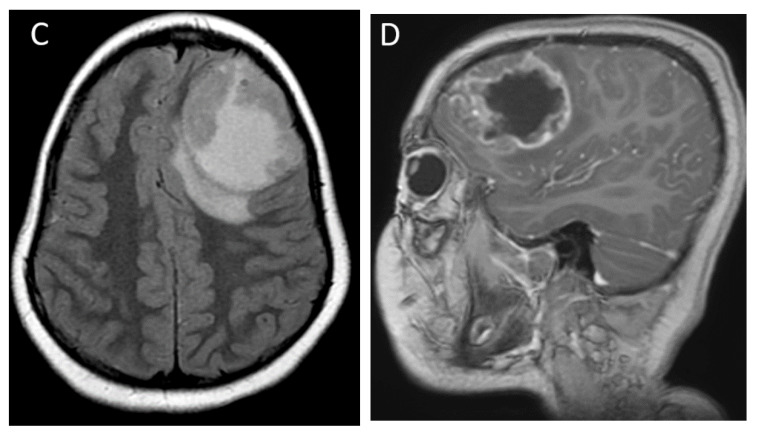
Figure 5.
A 6-month-old male with CNS embryonal tumor, NOS, WHO Grade 4. Axial DWI (A), axial SWI (B), axial T2 FLAIR (C) and sagittal post-contrast T1-weighted (D) images demonstrate a large mass in the left frontal lobe, with diffusion restriction of the peripheral solid components, scattered small amounts of blood products and/or mineralization, isointense T2 signal of solid portion, large area of central necrosis/cyst, and heterogenous post-contrast enhancement of the solid portions. Note the similarities in appearance to the HGG illustrated in Figure 1; however, this tumor presented at a much younger age.


