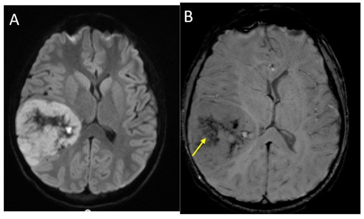
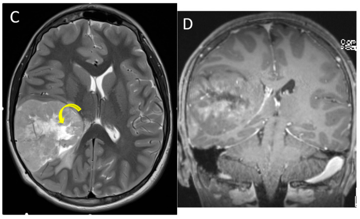
Figure 7.
A 14-year-old male with H3 G34R mutant high-grade glioma. Axial DWI (A), axial SWI (B), axial T2 (C) and coronal post-contrast T1-weighted (D) images demonstrate a large mass in the right parieto-temporal lobes, with diffusion restriction of the solid portions, central areas of blood products and/or mineralization (arrow), isointense T2 signal with central necrosis (curved arrow), and heterogenous post-contrast enhancement.


