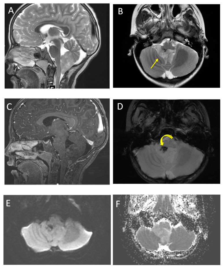
Figure 9.
A 6-year-old male with ependymoma. Sagittal and axial T2 images (A,B) demonstrate a heterogeneous lesion involving the fourth ventricle. Note the characteristic extension through the foramen of Luska (arrow). Post-contrast T1 imaging (C) demonstrates typical heterogeneous enhancement. SWI imaging (D) demonstrates a focus of low signal correlating with calcification (curved arrow). DWI (E) and ADC (F) imaging show increased ADC values of the tumor compared with adjacent parenchyma.

