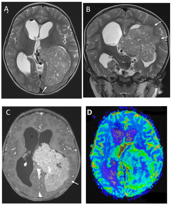
Figure 11.
A 2-year-old male with choroid plexus carcinoma. Axial and coronal T2-weighted (A,B), axial post-contrast T1-weighted (C) and axial perfusion rCBF (D) images demonstrate a large, predominantly solid mass with scattered tiny cystic foci, within the atrium and occipital horn of the left lateral ventricle. There is diffuse enhancement of the tumor and increased blood flow on the perfusion maps. The presence of parenchymal invasion (arrows on (A–C)) distinguishes choroid plexus carcinoma from papilloma.

