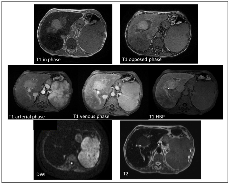
Figure 2.
MRI imaging of a 72-year-old female with secondary hemochromatosis, showing a nodular area in the IV segment. The area, adjacent to portal bifurcation, appeared as hyperintense in comparison to the surrounding liver parenchyma both in T1 in phase and opposed phase imaging. The liver parenchyma appears less hypointense in the T1 opposed phase (TE = 2.4 ms) than in the T1 in-phase image (TE = 4.8 ms) due to the iron overload. The lesion was also slightly hyperintense in T2 weighted images without restriction on DWI sequences. After the administration of contrast agent (Gd-EOB-DTPA), it seemed to have hyperenhancement on arterial phase without wash-out in venous phase. In the hepatobiliary (HBP), it appeared hyperintense. This lesion turned out to be a nodular area of iron sparing.

