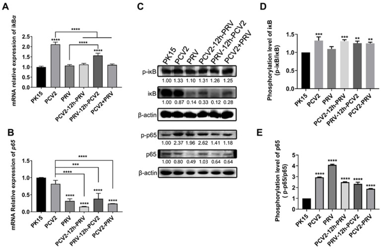
Figure 5.
Coinfection of PCV2 and PRV modulates the NF-κB signal pathway. (A,B) Expression levels of iκB and p65. The relative mRNA levels of iκB (A) and p65 (B) were examined using real-time PCR. The data are presented as the means ± SD. **, p-value < 0.01; ***, p-value < 0.001; ****, p-value < 0.0001. (C–E) Protein levels of p65 and iκB. Western blotting was conducted using P-IΚBα (Ser32 Ser36), IΚB-α, NFκB p65 Polyclonal Antibody, Phospho-NFκB p65 (Thr276) Polyclonal Antibody, and Anti-β-Actin Antibody as primary antibody, respectively. HRP-labeled Goat Anti-mouse IgG (H+L) and HRP-labeled Goat Anti-rabbit IgG (H+L) were used as the secondary antibody. β-actin was used as a control. The average expression level of the target protein in each group is shown below each lane. The protein amount of the PK-15 group is set to 1, and the values of other groups are the ratio with the PK-15 group. Unprocessed original images can be found in Supplementary Figure S4.

