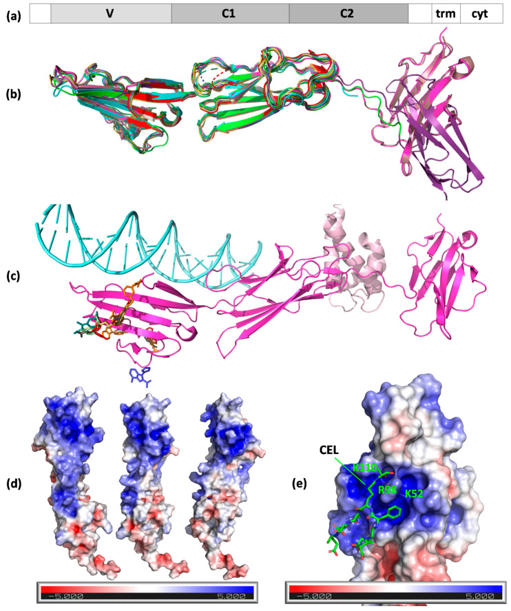
Figure 3.
Structures of RAGE and ligand binding mode. (a) The domain organization of the receptor: 1–21, signal peptide; 23–116, Ig-like V-type; 124–221, Ig-like C2-type 1; 227–317, Ig-like C2-type 2; 343–363, transmembrane helical; 364–404, cytoplasmic (disordered); (b) A chains (N chain in the case of 3o3u, gold) of RAGE structure models aligned to 3cjj A chains (VC1 fragment, green). Protein chains are shown as cartoons and are colored: 4oi7, cyan; 4lp5, dark violet; 4p2y, dirty pink; 4ybh, pink; 6xq1, yellow; 6xq3, orange; 6xq5, olive; 6xq6, pale green; 6xq7, dark cyan; 6xq8, red; 6xq9, gray; and 7lmw, dark blue. MBP fusion protein in 3o3u is not shown. A chains (N in the case of 3o3u) of RAGE aligned to 3cjj A chain (VC1 fragment) with low or very low RMSD (given in Å): 3o3u–0.991, 4lp5–0.889, 4oi7–0.759, 4p2y–0.544, 4ybh–0.602, 6xq1–1.094, 6xq3–1.221, 6xq5–1.122, 6xq6–1.107, 6xq7–1.161, 6xq8–1.325, 6xq9–1.241, and 7lmw–1.264; (c) RAGE VC1C2 (cartoon, PDB ID 4p2y, magenta) with its ligands (S100-A6, cartoon, light pink, 4p2y; DNA, light cyan, 4oi7) and inhibitors (colored sticks, from PDB IDs 6xq–6xq9 and 7lmw); (d) positively charged patch on the surface of RAGE (surface view, PDB ID 3cjj) shown in three orientations; (e) CEL peptide binding to the positively charged patch on RAGE surface (PDB ID 2l7u).

