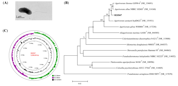
Figure 3.
Characterization of Agarivorans sp. B2Z047. (A) Transmission electron microscopy of cell of strain B2Z047. Cells were grown on MA at 30 °C for 48 h. Bar, 1 µm. (B) Neighbor-joining phylogenetic tree based on the 16S rRNA gene sequences, showing the relationships of strain B2Z047 (GenBank: OM278383) and the related species. Bootstrap values ≥ 70% (based on 1000 replications) were shown at branching nodes. Filled circles indicated that the corresponding nodes were also recovered in the trees generated with the maximum-likelihood and maximum-parsimony algorithms. Pseudomonas aeruginosa DSM 50071T was used as an out-group. Bar, 0.02 substitutions per nucleotide position. (C) Genome map of strain B2Z047. The circles from the inner to the outer: Circle 1 for GC content; and Circle 2 for GC skew.

