Figure 5.
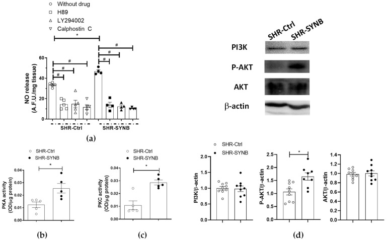
(a) Inhibitory effect of H89 (PKA inhibitor, 1 µmol/L), calphostin C (PKC inhibitor, 0.1 µmol/L) or LY 294002 (PI3K inhibitor, 10 µmol/L) on EFS-induced NO release in endothelium-denuded mesenteric rings from SHR-Ctrl and SHR-SYNB (n = 4–5 segments in each experimental group). Data (arbitrary fluorescence units/mg tissue) are expressed as mean ± S.E.M. * p < 0.05 SHR-Ctrl vs. SHR-SYNB (Student’s t-test). # p < 0.05 conditions without inhibitor vs. conditions with inhibitor in each group (Student’s t-test). (b) PKA activity, and (c) PKC activity in mesenteric arteries from SHR-Ctrl and SHR-SYNB (n = 5 segments from different animals in each group). Results (optical density (OD) units/µg protein) are represented as (mean ± S.E.M). * p < 0.05 (Student’s t-test). (d) Analysis for AKT and PI3K (P85 subunit) expression, and AKT phosphorylation at the T308 residue (P-AKT) in mesenteric arteries from SHR-Ctrl and SHR-SYNB (8–9 isolated arterial segments from different animals in each group). Lower panel: Densitometry analyses of the protein expression. Results (mean ± S.E.M) are expressed as protein expression relative to β-actin expression. * p < 0.05 (Student’s t-test).

