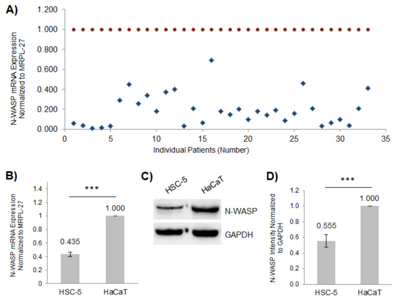
Figure 1.
Expression of N-WASP is reduced in SCC patient samples and the SCC cell line HSC-5. (A) The total RNA was extracted from the paraffin-embedded SCC and matched perilesional samples and reverse-transcribed to cDNA for quantifying the N-WASP expression relative to MRPL-27 using real-time PCR, normalized to individually matched perilesional samples (SCC values: blue diamonds, matched perilesional values: red circles); n = 33. (B) The total RNA was extracted from the HSC-5 and HaCaT cells and reverse transcribed to cDNA for quantifying N-WASP expression relative to MRPL-27 using real-time PCR, normalized to HaCaT cells. (C) Representative Western blots of N-WASP and GAPDH (loading control) in the HSC-5 and HaCaT cells; n = 3. (D) Densitometric quantification of the N-WASP/GAPDH ratio in HSC-5 cells normalized to HaCaT cells. All values are the mean ± SD, n = 3, *** p < 0.001.

