Figure 3.
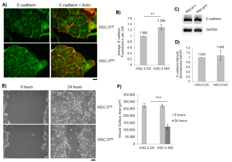
HSC-5NW cells have increased E-cadherin localization and reduced cell migration. (A) Representative immunofluorescence images of HSC-5CN and HSC-5NW cells stained for E-cadherin (green). Actin was stained with Alexa Fluor 568 phalloidin (orange–red). Scale bar represents 20 µm, n = 3. (B) Quantification of E-cadherin fluorescence from 20 randomly chosen cells per experiment based on the number of interacting cell–cell junctions and normalized to HSC-5CN cells. (C) Representative Western blots of E-cadherin and GAPDH in HSC-5CN and HSC-5NW cells; n = 3. (D) Densitometric quantification of the E-cadherin/GAPDH ratio in HSC-5CN and HSC-5NW cells normalized to HSC-5CN cells. Values are the mean ± SD, n = 3, p > 0.05. (E) Representative images of in vitro wounds of HSC-5CN and HSC-5NW cells at 0 and 24 h. Scale bar represents 50 µm, n = 3. (F) Quantification of wound areas performed in (E) at respective time-points using ImageJ. All values are the mean ± SD, n = 3, ** p <0.01, *** p < 0.001.

