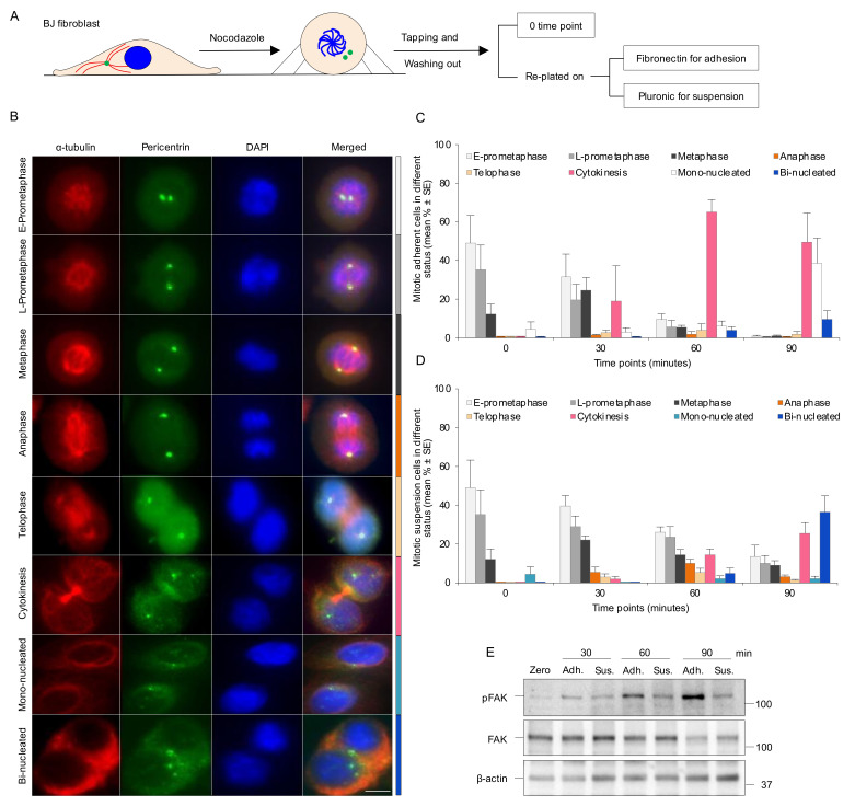
Figure 1.
Absence of integrin-mediated cell adhesion causes a delay in the early mitotic progression. (A) Experimental design illustrating how the isolated mitotic cells were used to check mitotic progression in the different culture conditions. Centrosomes, microtubules, and nucleus/chromosomes are shown in green, red, and blue, respectively. (B) Representative immunofluorescence micrographs illustrating the mitotic progression of BJ cells from early (E)- and late (L)-prometaphase to cytokinesis, the presence of divided cells (mono-nucleated cells), and the cells that failed to complete cytokinesis (bi-nucleated cells). The centrosomes and the mitotic spindle were labeled with antibodies against pericentrin (green) and α-tubulin (red) directly after nocodazole washout (0 min time-point) and after a 30, 60, and 90 min incubation period of the cells adhering to fibronectin or kept in suspension. Nuclei were stained with DAPI. Scale bar, 10 μm. (C,D) Mean (%) ± SE of the number of adherent (C) and non-adherent cells (D) in different mitotic stages determined based on centrosomes’ location, spindle formation, and nucleus status as shown in part B. The color of each bar on the graph corresponds to the different mitotic phases shown in the side color bar of part B. (E) Western blot of the mitotic cells treated as in (C,D) to monitor the phosphorylation of FAK at Tyr397 and total FAK at the described time points.

