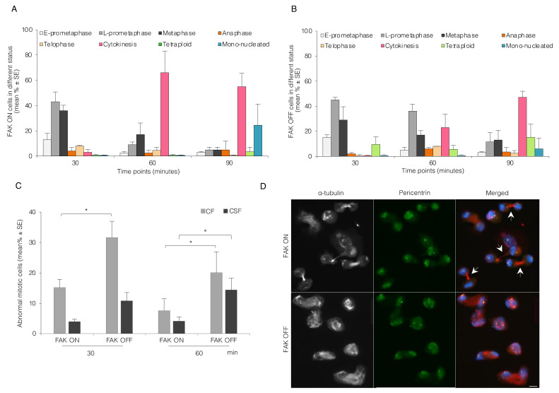
Figure 5.
Inhibition of FAK expression in Tet-FAK MEF cells impairs centrosome separation and bipolar spindle formation. (A,B) Mean (%) ± SE of the number of Tet-FAK ON (A) and Tet-FAK OFF cells (B) in different mitotic stages as described in Figure 1B. (C) Mean (%) ± SE of the number of Tet-FAK ON and OFF cells containing abnormal centrosomes (CF and CSF) present at different mitotic stages. (D) Representative immunofluorescence pictures illustrating FAK ON and FAK OFF cells 60 min after release from mitotic block. The arrows point to the microtubule bundle of cytokinetic cells (FAK ON). Pericentrin (green), α-tubulin (red/white), and DAPI (blue). Scale bar, 10 μm. p-values less than 0.05, 0.01, 0.001, and 0.0001 were shown by one star, respectively.

