Figure 6.

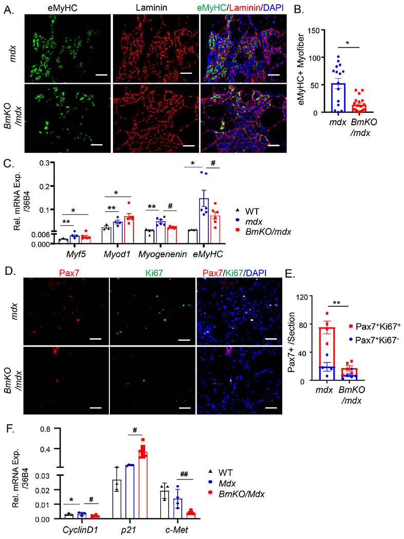
Bmal1 deficiency in mdx mice impairs regenerative repair by attenuating neo-myofiber formation and satellite cell proliferation. (A, B) Representative images of embryonic myosin heavy chain (eMyHC, green), laminin (red) and DAPI (blue) in TA from mdx and BmKO/mdx mice (A), and the quantification of fibers with eMyHC expression as a percentage of total fibers from mdx and BmKO/mdx TA cross sections (B). Scale bar: 50 μm. (C) RT-qPCR analysis of myogenic factor expression in TA of WT (n=3), mdx mice (n=7) and BmKO/mdx mice (n=4). (D, E) Representative images of Pax7 (Red), Ki67 (Green) and DAPI in TA sections from mdx (n=4) and BmKO/mdx mice (n=5, D), and quantification of Pax7+ myofibers with or without Ki67 co-staining in TA cross sections (E). Scale bar: 50 μm. (F) RT-qPCR analysis of key factors involved in satellite cells proliferative response in TA muscle of WT (n=3), mdx mice (n=7) and BmKO/mdx mice (n=4). *, ** P≤0.05 or 0.01 vs. WT, #, ##: P≤0.05 or 0.01 BmKO/mdx vs. mdx mice.
