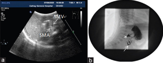
Figure 1.

(a) Ultrasound image showing the reverse positional relationship between the superior mesenteric vein and superior mesenteric artery. The superior mesenteric vein is located at the left front of the superior mesenteric artery. (b) Upper gastrointestinal imaging examination showing the corkscrew sign in the duodenum, indicating a spiral curve similar to “apple-peel/twisted ribbon” formed due to midgut volvulus
