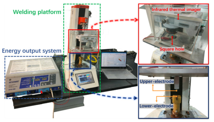
Figure 1.
Physical map of H.F.E.W. experimental setup. In the experiment, the rigid structure composed of a tensimeter base, infrared thermal imager and upper-electrode compresses the tissue downward, the infrared thermal imager can record the temperature distribution in the welding process through the square hole, and the electrical parameters are measured and displayed by an oscilloscope (including current ring and voltage probe) (not shown).

