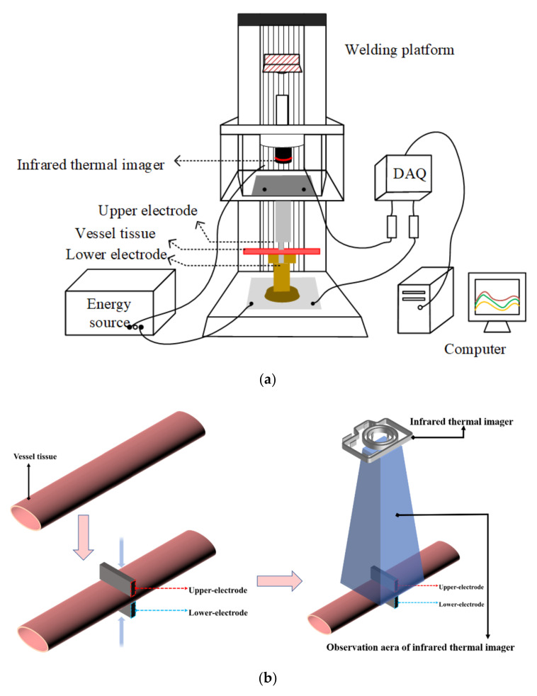
Figure 2.
The treated vessel tissue is placed between the upper and lower electrodes, and sufficient pressure is applied to fix it. After the pressure is stabilized, the upper and lower electrodes deliver energy to the vessel tissue, and the infrared thermal imager begins to record the tissue temperature until the end of the welding experiment. (a): Schematic diagram of H.F.E.W. experimental setup. (b): Flow chart of vessel tissue welding experiment.

