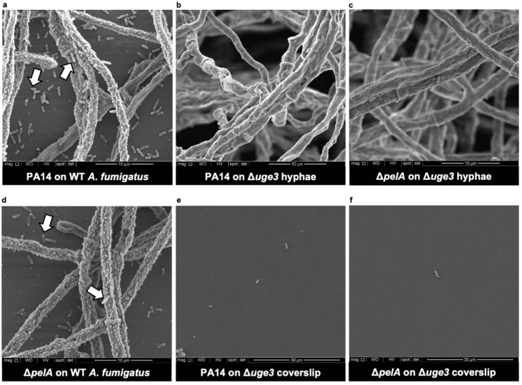
Figure 3.
GAG mediates the adherence of P. aeruginosa to A. fumigatus hyphae. Scanning electron microscopy images of co-cultures of wild-type P. aeruginosa (PA14) with (a) adherent wild-type (WT) A. fumigatus hyphae on coverslips or (b) non-adherent GAG-deficient A. fumigatus hyphae (Δuge3). Co-cultures of Pel-deficient P. aeruginosa (ΔpelA) with (c) non-adherent GAG-deficient A. fumigatus hyphae (Δuge3) or with (d) adherent wild-type A. fumigatus hyphae (WT) on coverslips. (e) Coverslip of (b) and (f) coverslip of (c). Representative images of 2 independent experiments. (a–d) Imaged at 10,000×. (e,f) Note a lower magnification at 5000×. White arrows indicate adherent bacteria to hyphae and coverslips in the interstitial spaces between hyphae.

