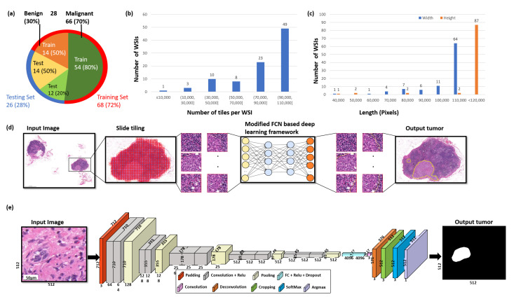
Figure 2.
The overview of the data and the proposed deep learning framework presented in this study. (a) Distribution of data between malignant and benign samples and for training and testing data, respectively. (b) The number of tiles delivered per WSI. (c) The width and height distributions of the WSIs are shown in blue and orange, respectively. (d) The proposed deep learning framework for the segmentation of breast cancer. Firstly, Otsu’s method is used to threshold the slide image to efficiently discard all background noise. Secondly, each WSI is formatted into a tile-based data structure. Thirdly, the tiles are then analyzed by a deep convolutional neural network to produce the breast cancer metastasis segmentation results. (e) Illustration of the proposed modified FCN architecture.

