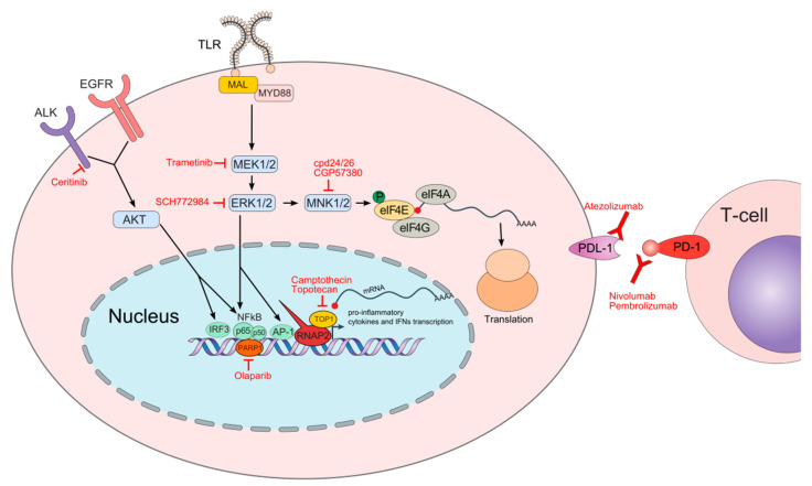
Figure 1.
Schematic depiction of molecular pathways and processes involved in cellular responses upon pathogen infection and respective oncology drugs repurposed for experimental sepsis treatment. The names of oncology drugs discussed herein are in red font next to their molecular targets. Following toll-like receptor (TLR) activation, the myeloid differentiation primary response protein 88 (MYD88) together with MYD88 adaptor-like protein (MAL) are recruited to the TLRs initiating the cascade of molecular events activating mitogen-activated protein kinase (MAPK) components including MEK and extracellular signal-regulated kinase (ERK) that ultimately mobilize chromatin recruitment of interferon regulatory factors (IRFs), nuclear factor (NF)-κB, and activator protein 1 (AP-1) to gene loci initiating expression of the specific immune responses. ERK also indirectly influences translation by regulating MAPK-interacting kinase (MNK) that phosphorylates the eukaryotic translation initiation factor 4E (eIF4E) at Ser209 from a cap-binding complex leading to the translation of transcripts encoding pro-inflammatory cytokines, including tumor necrosis factor (TNF)α. Anaplastic lymphoma kinase (ALK) participates with the epidermal growth factor receptor (EGFR) to promote AKT stimulation, which then activates NF-kB and IRF3 factors to induce the expression of proinflammatory cytokines and interferon β (IFN β). In the nucleus, the poly (ADP-ribose) polymerase 1 (PARP1) acts as a transcriptional co-regulator of the NF-κB transcriptional factor while the topoisomerase 1 (TOP1) facilitates polymerase 2 RNA (RNAP2) recruitment to the genes encoding pro-inflammatory mediators. Checkpoint proteins including programmed cell death protein 1 (PD-1) and PD-1 ligand (PDL-1) play an essential role in transitioning from a hyper- to hypo-inflammatory response. Both PD-1 and PDL-1 are expressed on immune cells while PDL-1 is also expressed on non-immune cells.

