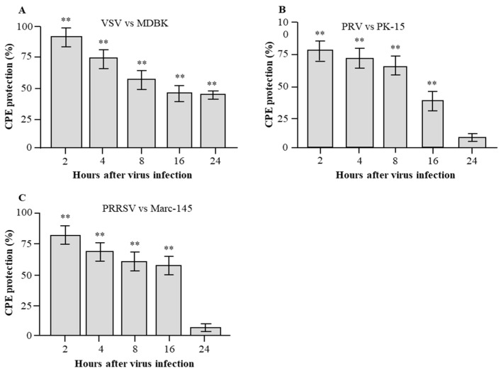
Figure 6.
Detection of post-infection antiviral activities of rPoIFN-β. Three cell lines MDBK cells (A), PK-15 cells (B) and Marc-145 cells (C) were infected with the indicated viruses VSV (A), PRV (B) or PRRSV (C) and then treated with a fixed concentration of rPoIFN-β. At different time points post infection, the post-infection antiviral activities were measured in triplicates by CPE inhibition assay. ** indicates significant difference (p < 0.01) in CPE protection of rPoIFN-β as compared with the virus control (VC).

