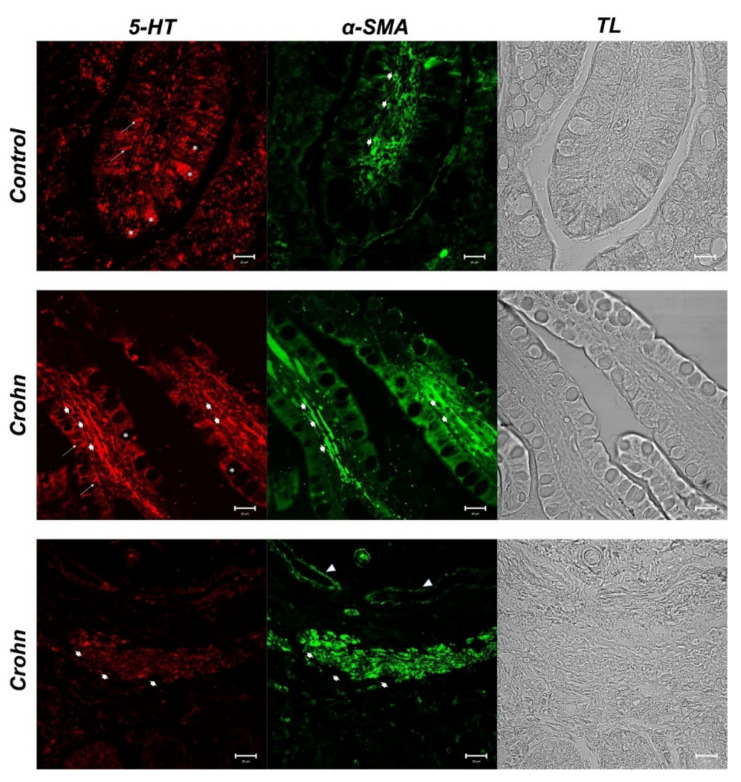
Figure 2.
Human intestine. 5-HT and α-SMA, 20×, scale bar 20 nm. In healthy intestine sections, red fluorescence evidenced serotonergic enteroendocrine cells (arrows) and the goblet cell cytoplasm (*). The positivity to α-SMA was observed in stromal cells (fibroblasts and mesenchymal cells) (large arrows). In CD samples, enterochromaffin cells (ECs), positive to 5-HT, were evident in the epithelium (arrows). Lamina propria cells with a high positivity to 5-HT were detectable (big arrows). The goblet cell cytoplasm was 5-HT negative (*). α-SMA positive cells in the lamina propria below the epithelium (large arrows). Blood vessels (arrowhead). The micrographs are also equipped with transmitted light (TL) to visualize the organ morphology.

